Pneumothorax
A Pneumothorax is an abnormal collection of air or gas in the pleural space separating the lung from the chest wall. This buildup of air puts pressure on the lung, so it cannot expand as much as it normally does when you take a breath.
Classfication:
A. Spontaneous Pneumothorax
Spontaneous pneumothoraces are a common condition, which affects thousands of individuals throughout the world each year.
Spontaneous pneumothoraces are divided into two types:
(i) Primary, which occurs in the absence of known lung disease, and (ii) Secondary, which occurs in someone with an underlying lung condition.
 The exact cause of primary spontaneous pneumothorax is unknown, but established risk factors include being male, smoking, and having a prior history of a spontaneous pneumothorax or a family history of pneumothorax. In most cases, primary spontaneous pneumothorax results from the rupture of blebs (air-filled sacs on the lung). This causes air to escape from the lung and enter the chest cavity causing the lung to collapse.
Secondary spontaneous pneumothorax occurs in the setting of a variety of lung conditions. The most common of which is chronic obstructive pulmonary disease (COPD), which accounts for approximately 70% of cases. Other lung diseases that may significantly increase the risk for pneumothorax include asthma, cystic fibrosis, tuberculosis, whooping cough and lung cancer to name a few.
A spontaneous pneumothorax has a very high rate of recurrence. It is estimated that the probability of recurrence is as high as 50% in some patients.
B. Tension Pneumothorax
Occasionally, the amount of air in the chest increases markedly when a one-way valve is formed, leading to a tension pneumothorax. As the air escapes from the lung and enters the pleural space, increased pressure can cause a mediastinal shift, in which the heart, trachea, and great vessels are pushed towards the opposite side. This condition is a medical emergency that can cause steadily worsening oxygen shortage and low blood pressure. Unless reversed by immediate effective treatment, it can progress rapidly resulting in death.
C. Traumatic Pneumothorax
A traumatic pneumothorax occurs when some physical trauma causes a pneumothorax. Any physical injury such as a knife wound or high impact injury can result in a pneumothorax. Medical procedures such as transbronchial biopsy, thoracentesis, chest tube insertion, etc., can all increase the risk of a pneumothorax.
Symptoms:
Symptoms of a pneumothorax include sudden onset of chest pain on the side of the pneumothorax. The pain is sharp and may lead to feelings of tightness in the chest. Shortness of breath, rapid heart rate, rapid breathing, cough, and fatigue are other symptoms of pneumothorax. The skin may develop a bluish color (termed cyanosis) due to a decrease in blood oxygen levels.
Diagnosis:
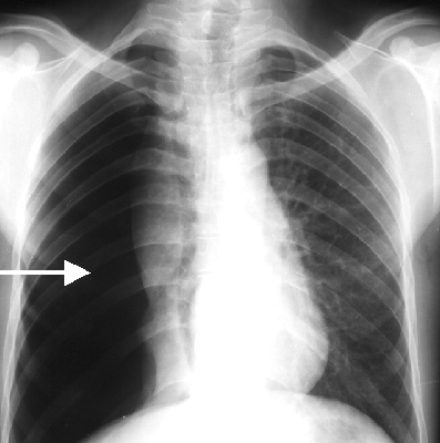
Examination of the chest with a stethoscope reveals decreased or absent breath sounds over the affected lung. There may be shifting of the trachea and mediastinal structures to the opposite side. Diagnosis of a pneumothorax by physical examination alone can be difficult or inconclusive (especially if the pneumothorax is small), so a chest X-ray or computed tomography (CT) scan is usually used to confirm its presence.
Treatment:
A small pneumothorax typically resolves without treatment and require only monitoring. In larger pneumothoraces, or when there are marked signs and/or symptoms, the air may be extracted with a syringe or a chest tube connected to a one-way valve system. Occasionally, surgical interventions are required when tube drainage is unsuccessful, or as a preventative measure, if there have been repeated episodes.
Surgical treatments usually involve pleurodesis (which induce the layers of pleura to stick together) or pleurectomy (the surgical removal of pleural membranes).
Further Reading
The article above is meant to provide general information and does not replace a doctor's consultation.
Please see your doctor for professional advice.