Kidney Stones
 The urinary tract is the system that produces urine and carries it out of your body. It is made up of the kidneys, the tubes that connect the kidneys to the bladder (the ureters), the bladder, and the tube that leads from the bladder out of the body (the urethra).
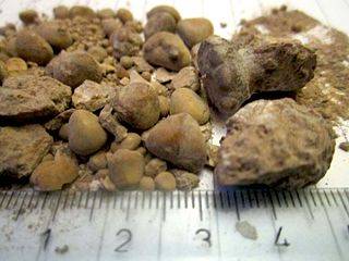
Sometimes, stones may form along the urinary tract and give rise to problems as a result of obstruction to urinary flow. The most common type of stone contains calcium oxalate or calcium phosphate. Less common types of stones include uric acid stone (associated with gout), and cystine stones.
What Causes Kidney Stones?
Kidney stones form when there is a high concentration of certain chemicals in the urine (eg. calcium, phosphate, oxalate and uric acid) on one hand, and a low concentration of substances that prevent stone formation (the "inhibitors" such as citrate and magnesium) on the other.
Certain conditions affecting the urinary tract, such as urinary tract infections, cystic kidney diseases as well as certain metabolic diseases, are linked to stone formation. Calcium oxalate stones are also more likely to develop in people with chronic inflammation of the bowel, or in those who have had intestinal bypass surgery.
Kidney stones may also be an inherited disease. If other people in your family have had kidney stones, you may have them too.
What Are The Symptoms?
 Most stones that remain in the kidneys do not cause any symptoms. It is when the stone moves downward into the ureter, blocking the flow of urine, that the first symptoms suddenly appear:
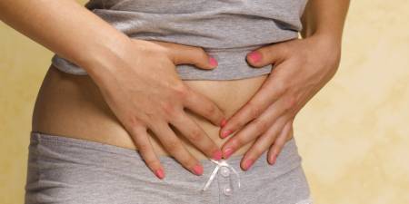
• Sudden, severe pain that gets worse in waves. The pain is typically felt in the lower back (in the loin region around the area of the kidneys) and may shift to the lower abdomen and groin. People who have had a kidney stone often describe the pain as "the worst pain they have ever had."
• Often, there is associated nausea and vomiting.
• As the stone moves through the urinary tract, blood may appear in the urine (known as haematuria).
• As the stone moves down the ureter closer to the bladder, there may be the sensation of needing to urinate more frequently, and with greater urgency. Some may also develop a burning sensation during urination (dysuria). These symptoms of frequency, urgency and dysuria and known as the "lower urinary tract symptoms". Lower urinary tract symptoms associated with a fever is more likely to be due to a urinary tract infection rather than stones.
What Are The Risk Factors?
Several factors may increase your likelihood of developing kidney stones.
Risk Factors You Can Control:
• How much fluid you drink. The most common cause of kidney stones is not drinking sufficient water. You can tell if you drink enough fluids by observing the colour of your urine - which should be clear or with a tinge of yellow. If your urine appears dark in colour, it means that you are not drinking enough.
• Diets high in protein, sodium, and oxalate-rich foods, such as dark green vegetables, increase your risk for kidney stones. Depending on the type of stones you have, diet may or may not be a factor.
• Being overweight can increase insulin resistance and increase calcium in the urine.
• Consuming Medication. Certain medication can increase the likelihood of kidney stone formation.
Risk Factors You Cannot Control:
• Age and gender: Stones occur more frequently in men, and most commonly between 40 to 70 years old. Postmenopausal women and women who have had their ovaries removed are also at increased risk.
• A family history of kidney stones.
• History of frequent urinary tract infections.
• Presence of other diseases, such as hyperparathyroidism, or gout.
• Intestinal surgery or gastric bypass surgery.
Diagnosis:
Diagnosis of kidney stones is made on the basis of information obtained from the history, physical examination, urinalysis, and radiographic studies.
 • A plain film abdominal X-ray (KUB) gives a picture of the kidneys, the bladder, and the tubes that connect the kidneys to the bladder (ureters). Radio-opaque stones may be visible on plain X-rays.
• Ultrasound of the kidneys and bladder.
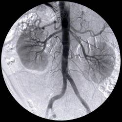
• Intravenous Urogram (IVU) is a contrast study which can identify stones in the urinary tract with much greater accuracy compared to standard X-rays.
• Computerised tomography (CT) scan.
• Additional urine and blood tests may be required to exclude a urinary tract infection or impairment of kidney function.
• Stone analysis. You may be asked to collect stones by straining your urine through a strainer or gauze.
• Urine collection for 24 hours, to measure volume, pH, calcium, oxalate, uric acid, and other substances that may have caused the stone to form.
Prevention:
Your chances of developing another stone are increased if you have had more than one kidney stone. Therefore, prevention is important.
Adequate Water Intake: Try to drink about 8 to 10 glasses of water a day. You will know that you are drinking enough water when your urine is clear or light yellow. If it is dark yellow, it means that you are not drinking enough. If you have kidney, heart, or liver disease and have fluid restrictions, talk with your doctor before increasing your fluid intake.
Calcium Intake: In the past, people who had calcium stones were advised to avoid dairy products and foods high in calcium content. Recent studies however, have found that foods high in calcium (including dairy products), may in fact help to prevent calcium stones.
Oxalate Intake: Limiting consumption of foods containing high amounts of oxalate (such as spinach, strawberries, nuts, wheat germ, dark chocolate, cocoa, black tea, liver, marmalade and sweet potatoes)
Meat Intake: If your urine is highly acidic, you may need to reduce your consumption of meat, fish and poultry, as these foods increase in the amount of acid in the urine.
Treatment:
Both the size as well as the position of the stone influence the rate of spontaneous stone passage. For instance, up to 98% of small stones (ie. less than 5 mm in diameter) may pass out spontaneously within 4 weeks. For larger stone (5 - 10 mm in diameter), the rate of spontaneous passage drops to about 50%. Hence, for small stones, most people don't need any treatment other than taking pain medicine and drinking enough fluids.
Initial stone location also influences the likelihood of spontaneous stone passage. Stones located in certain parts of the urinary tract are more likely to pass out spontaneously.
{loadpostion linead}
Medical Therapy:
Certain medication may help prevent calcium and uric acid stones from forming, by controlling the amount of acid or alkali in the urine, which are key factors in stone formation. The type of medication will depend on the type of stones you develop. Allopurinol may be useful in some people who form uric acid stones.
Surgical Therapy:
1. Extracorporeal Shock Wave Lithotripsy (ESWL)
ESWL uses shock waves that pass easily through the body but are strong enough to break up a kidney stone. The stones are broken down into smaller particles that pass easily through the urinary tract in the urine. If the stone is not completely shattered with one treatment, additional treatments may be required. ESWL is generally done on an outpatient basis and recovery time is relatively short.
Patients may experience some pain on urination (as the small stones are passed out of the urinary tract) as well as some blood in the urine.
2. Percutaneous Nephrolithotomy or Lithotripsy (PCNL)
PCNL is used when the stone is large or in a location that is not well suited for the use of ESWL.
The surgeon makes a small incision in the patient's back, then puts a hollow tube into your kidney and either removes (lithotomy) or breaks up and removes (lithotripsy) the stone. A small tube known as a nephrostomy tube is left in the kidney for a few days.
3. Uretheroscopic Stone Removal
This may be needed for mid to lower ureteric stones. The surgeon passes a ureteroscope (a small fibreoptic instrument) through the urethra, into the bladder and then then ureter, to locate and fragment the stone. The fragmented pieces of stone is then removed. A small stent may be left in the ureter for awhile to help with the flow of urine.
The article above is meant to provide general information and does not replace a doctor's consultation.
Please see your doctor for professional advice.