Osteoarthritis
 Osteoarthritis is the most common type of arthritis, occurring in up to about 10% of adults, with as many as 50% of the elderly suffering from it. It is basically a degenerative form of arthritis, in which the cartilage, whose function is to cushion the joints, gets worn out with age.
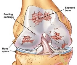
This “wear-and-tear” of the cartilage over time, results in the bone surfaces becoming less protected and increases friction between the bones during movement. This friction eventually results in pain, swelling and loss of mobility. In more advanced stages, the joint loses its normal shape and bony spurs may grow on the edges of the joint. Bits of bone or cartilage may break off and float inside the joint space, further causing pain and loss of mobility.
CAUSES & SYMPTOMS
The cause is multi-factorial, but the following would increase your risk:
• Being overweight
• Getting older
• Previous injury to the joint
• Mechanical stresses on the joint from high impact sports, certain jobs, pathological or congenital mal-alignment of bones
Symptoms in the initial stages may include pain, tenderness, stiffness, creaking and locking of the affected joint. As the arthritis progresses, there may be swelling of the joint due to collection of synovial fluid within the joint. In the more advanced stages, there is bony deformity (caused by bony spurs) and mal-alignment of the limb (eg. “varus” deformity of the knee). Patients experience increasing pain upon weight bearing, thus limiting walking, and ultimately, even standing.
Osteoarthritis commonly affects the hands, feet, spine and weight-bearing joints, such as the hips and knees. In the smaller joints, such as in the fingers, hard bony swellings called Heberden's nodes and Bouchard's nodes may form. These are typically not painful, but they do limit joint movement.
DIAGNOSIS & MANAGEMENT
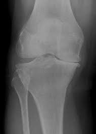
Diagnosis can often be made by your doctor with reasonable certainty by a thorough physical examination. X-rays are used to confirm the diagnosis as well as to  document progressive X-ray changes (thinning of cartilage, bony spurs, loose bodies, mal-alignment of joint etc) as the condition progresses.
Non-Pharmacological Management:
• Weight loss – Excess body weight puts more strain on the knee joints. A typical vicious cycle exists: (1) Overweight person develops knee osteoarthritis (2) painful knees reduce mobility (3) with reduced mobility, more weight is gained (4) more weight worsens the arthritis.
• Regular exercise – regular aerobic, strengthening and range of motion exercises help strengthen muscles that stabilize the joints,
• Adequate intake of Calcium and Vitamin D for bone strength.
• Warm soaks and heat packs to help relief pain.
• Avoid excessive walking during periods of acute pain.
• Orthoses and walking aids – splints and braces help with joint alignment and weight redistribution. Walking frames and crutches help take load away from the arthritic knee.
• Physiotherapy
• Acupuncture
Pharmacological Measures:
• Pain-killers – paracetamol-based medication
• Non-steroidal anti-inflammatory drugs (NSAIDS) etc.
• Glucosamine and/or chondroitin sulfate.
• Topical rubs with NSAIDS or capsaicin.
• Intra-articular joint injections by a doctor
Surgical Treatment:
• Joint lavage (wash out) and arthroscopic debridement (clearing)
• Osteotomy – a wedge of bone located near the damaged joint is removed to realign the knee. This causes a shift of weight from the area of damaged cartilage to the area where there is more healthy cartilage.
• Total Joint Replacement – considered to be the last resort option in which the severely arthritic joint, having failed more conservative methods of therapy, is replaced with a prosthetic joint.
Further Reading
The article above is meant to provide general information and does not replace a doctor's consultation.
Please see your doctor for professional advice.
